Retinal Fundus Images, Ground Truth of Vascular Bifurcations and Crossovers
The vasculature of the eye can provide amazing insight into systemic cardiovascular health. For those studying the vasculature of the eye as an indicator of pathophysiological states, normative ground truth datasets become critically important. Recently, George Azzopardi and Nicolai Petkov have made their annotated database of ground truth imagery of vascular bifurcations and crossovers in …
Continue reading “Retinal Fundus Images, Ground Truth of Vascular Bifurcations and Crossovers”
Notable Paper: Localization of Melatonin Receptor 1 in Mouse Retina and Its Role in the Circadian Regulation of the Electroretinogram and Dopamine Levels
I’ve watched the development of the circadian rhythm research starting with Joe Takahashi‘s work discovering CLOCK in the mammalian SCN back in 1994. Since then there has been an explosion of circadian rhythm biology work including an I suppose, unsurprising amount of research in the retina proper. This paper by Anamika Sengupta, Kenkichi Baba, Francesca Mazzoni, Nikita V. Pozdeyev, Enrica …
Rattlesnake!
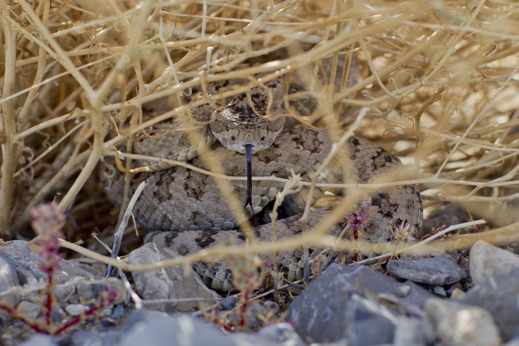
Utah, West Desert
Iris Tumor
This image demonstrates an iris tumor that is sitting in the angle. Photograph was made by James Gilman of the Moran Eye Center using a Goldmann 3-mirror lens scatter illumination with a Zeiss photo slitlamp and a Nikon D-1X camera.
Bassam’s B-Day

Notable Paper: Structural basis of PIP2 activation of the classical inward rectifier K+ channel Kir2.2
The inward rectifying K+ channels (IRKs) are common ion channels that encompass seven distinct subtypes, each a potential target for pathology or a potential actor in various insults to the nervous system, the retina included. IRKs in the retina are found in Müller glia, retinal pigment epithelium and on neurons in retina, so are fundamental to retinal …
Flying… and a Soapbox.

Portraits of Looks by Stefano da Luigi
This project called “Bianco, a multimedia project on visual deficiencies” by an amazing photographer/photojournalist Stefano da Luigi was sent by photojournalist extraordinaire and friend of Webvision, Trent Nelson. Stefano da Luigi’s work in this photoessay, a winner of the Visa Pour L’Image, Perpignan 2011 Multimedia Award is a stunningly intimate series of images of people experiencing …
Notable Paper: Neural Organization and Visual Processing in the Anterior Optic Tubercle of the Honeybee Brain
I have a certain fondness for insects and believe that our understanding of vision and visual pathways can benefit greatly from the study of insect visual systems. Our understanding of visual processing is actually pretty limited and simpler visual systems to study from eye to brain are found in insects compared to vertebrates. In …
Notable Paper: The Newly Sighted Fail to Match Seen With Felt
I missed this paper in the chaos and runup to ARVO, but its conclusions are remarkably compelling. Imagine that you were blind for years, perhaps from birth and suddenly, you were able to see with perfect clarity. Would you be able to recognize items like a pyramid, a box or a sphere by sight when …
Continue reading “Notable Paper: The Newly Sighted Fail to Match Seen With Felt”
Tour de Utah

Toxic Anterior Segment Syndrome (TASS)
Photographer: Paula F. Morris, CRA, FOPS Moran Eye Center. Diffuse and tangentially illuminated images of corneal deposits and folks in TASS Syndrome. Images taken a using a Zeiss clinical slit-lamp biomicroscope with a Nikon D1S digital back at 24 X and 40 X. TASS or Toxic Anterior Segment Syndrome is an inflammation of the anterior segment of …
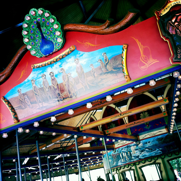
Carousel