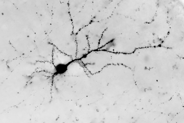
I am pretty tickled that this technique worked and am loving this small portion of data. I cannot say more than this because it is derived from data that we are helping some other folks out with, but this image was assembled with techniques that I wanted to try on another project some time ago. Unfortunately for that group, those people did not listen and I never ran the experiment for them. Today however, a more receptive audience came by asking how to solve this particular problem. I instructed them on how to collect the data for the experiment which took about 2 hours. After collecting the data, we had this image (part of a much larger image) in less than 2 minutes. It works and it works exceedingly well by taking a 3D problem and reducing it to a pseudo 2.5D problem (or 2.1D problem as Robert amusingly noted today).

You rock.
Thanks boss.
That’s amazing…
Thanks Mike. Check out all the dendritic spines on this neuron. Its neat that they showed up as well as they did.
Just think all those million of neurons firing, working together at the speed of light….so people can post cat pics on the internet….
Ha! Totally. I love LOL cats.
this is incredible!
Muchas gracias Scott.
beautiful!
is that a single filled cell? outside of the center of the image, are those axonal boutons? (tell me more!)
This is a section of visual cortex that has immunoprobes directed against a specific target. I cannot say more as it is from a colleagues dataset that we are helping to collect data for their grant. The boutons you are seeing are actually spines from that particular neuron.
What are the cells here other than the dendritic cells?
Hey Jack. This is a cortical neuron transfected with GFP. There are other cells here that are invisible in this image that are glial cells.