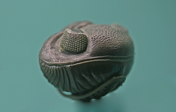
Trilobites were one of the most successful marine arthropods that lived from the Early Cambrian throughout the Devonian, finally going extinct in the Permian ages, a run of over 270 million years. They are well represented in the fossil record and even the earliest forms had complex compound eyes much like modern arthropods. These eyes had elongated lenses composed of calcite that modeling has revealed to provide excellent optical properties with good depth of field and little to no spherical aberration. These lenses brought light to photoreceptor cells at the base of the lens, but we’ve never before had an understanding of what that structure or anatomy looked like.
The problem of course with the fossil record is that very little internal structure remains in fossilized specimens. However, a very cool new study that examines trilobite eyes through X-ray tomography by Brigitte Schoenemann and Euan N. K. Clarkson reveals how these cells looked, down, perhaps to the cellular level. Followup work with μct-scanning and synchrotron radiation analysis reveals that the sensory structures (like rod or cone outer segments) are arranged in flower petal like structures around a central, diamond shaped photoreceptor cell body with pigment granules packed in-between. Its kind of like a modern limulus eye (image here).
It will be interesting to see if they can image other species of trilobite to get an evolutionary look at how eyes and perhaps primitive retinas developed over 500 million years ago.