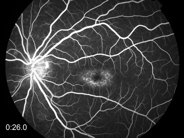
Venous phase fluorescein sodium angiogram of a dark choroid in Stargardt’s disease or fundus flavimaculatus. Stargart’s Disease is a progressive retinal degenerative disease caused by mutations in one of 3 genes, ABCA4, ELOVL4 or PROM1. The disease is a form of juvenile onset macular degeneration affecting the retinal pigmented epithelium (RPE). The disease onset and progression is usually bilateral.
Fundus autofluorescence is generally accepted as a standard means of determining the extent of RPE atrophy in Stargart’s disease as well as fluorescein examination of the vasculature. However, fluorescein angiography is an additional examination designed to image the blood flow behind the retina in the choroid. This image above shows mottled fluorescence in the macular region suggesting that there are abnormal regions of circulation present in the choroid behind the RPE of the macula.
50 ° image was made by Paula F. Morris, CRA, FOPS of the Moran Eye Center with a Zeiss FF 450+ fundus camera with Escalon digital capture system