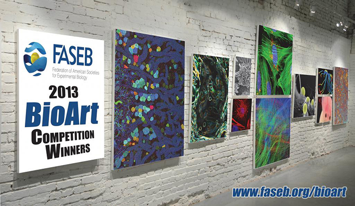
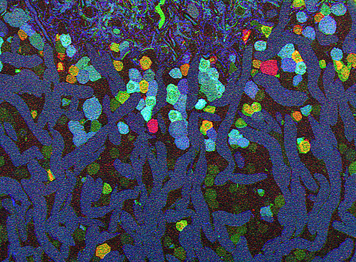
Robert Marc and I just found out that this image above was one of the 2013 FASEB BioArt Winners (Press release here). This image appeared here a little while ago and shows a region of an amazingly complex retina from a goldfish (Carassius auratus auratus) analyzed using tools called Computational Molecular Phenotyping (CMP) that reveal the metabolic state of the all cell types in tissues. These cells were labeled with antibodies for the presence of two fundamental amino acid metabolites (anti-glycine in red, anti-GABA in blue) and an amino acid tracer of physiologic activity (anti-AGB in green). These labels allow us to visualize the metabolic state and therefore, classes of bipolar, amacrine and horizontal cells.
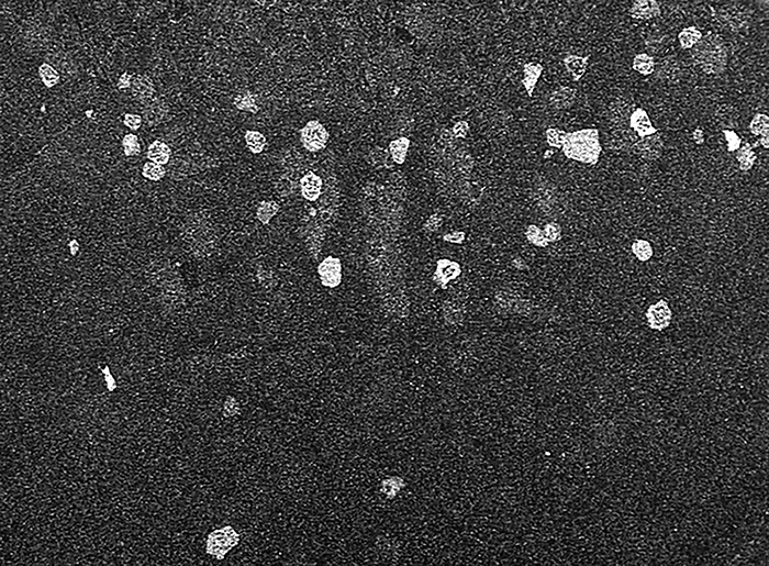
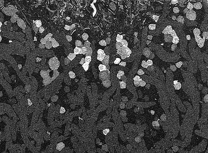
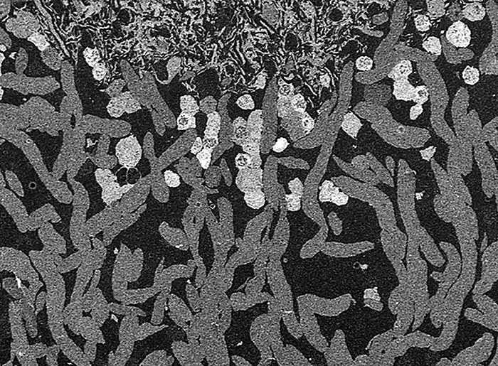
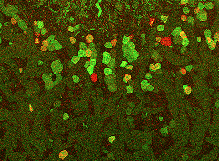
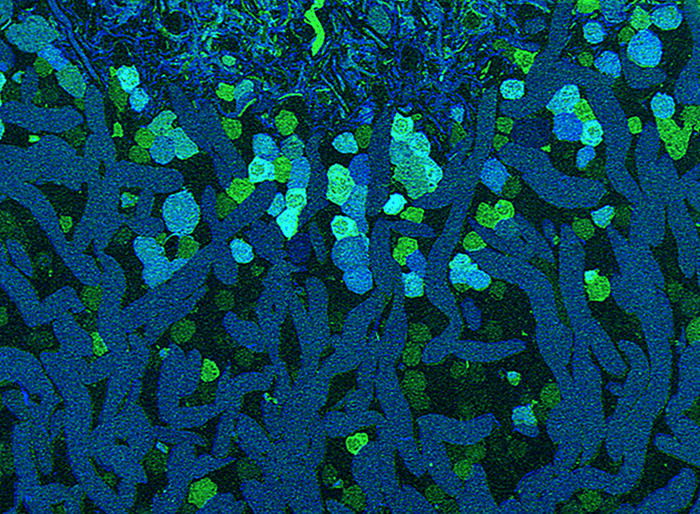
The individual labeled images are below in grayscale along with a couple of two color composites. Note that these are just 3 color images. The whole point behind CMP is that we can do this in N-space or use N labels to look at co-segregation of small molecular signals. We commonly look at 7-12 probes at once for instance, but they are difficult to show in traditional rgb images.
Glycine
AGB
GABA
Glycine and AGB in red and green.
AGB and GABA in green and blue.
The NIH National Eye Institute provided support for this research project that seeks to map retinal networks from both normal and diseased tissues like retinitis pigmentosa and age-related macular degeneration. FASEB or the Federation for American Societies for Experimental Biology is a coalition of biomedical researchers that works to advance health and promote progress and education in biological and biomedical sciences. The BioArt competition was designed to help communicate that work through compelling imagery and we are grateful that FASEB has found this image to meet that standard.
A 1000px wide image can be downloaded here.
Congrats !
Thanks man. When is the next coffee morning?