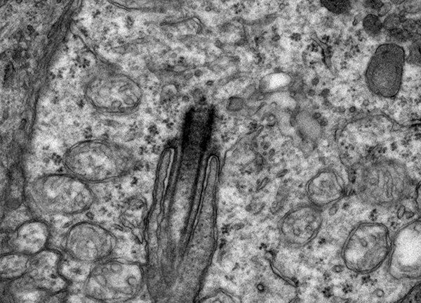
With the exception of a few types of cells, (acinar cells, T lymphocytesand hepatocytes), every cell in your body has a cilia. In the vision community, we are used to seeing these structures in the distal portion of the photoreceptors. The reality is that every cell in the retina has a cilium and some cells use the cilia as a means to expand a very specialized function like the photoreceptor outer segment or the hair cell or the respiratory epithelium of the lung. This particular cilia was found in an amacrine cell in a rat retina.
Cilia were thought for a long time to be vestigal organelles that are formed in development, then left over after the developmental process ended. Prachee Avasthi Crofts in the Wallace Marshall laboratorynotes that “cilia are signaling centers capable of sensing a variety of extracellular stimuli: fluid flow in the kidney, odorants in olfactory neurons, and hormones in the satiety center of the brain. Motile cilia in the trachea and brain ventricles can also generate flow of mucus and cerebrospinal fluid respectively. Dysfunction in conserved ciliary structure and function therefore results in a variety of disorders (termed ciliopathies) which include polycystic kidney disease, anosmia, obesity, bronchiectasis and hydrocephalus, to name a few.
In the retina, the outer segments of photoreceptors that sense light are in fact modified sensory cilia with conserved mechanisms of formation and maintenance. Thorough characterization of phototransduction proteins that reside in the outer segment as well as rapid turnover of outer segments to recycle spent membrane and protein make this system an excellent model to study cargo transport within cilia. Furthermore, a hallmark of many pleiotropic ciliopathies is retinal degeneration that results from abnormal photoreceptor cilia function. Investigation of photoreceptor cilia dysfunction can yield much insight into generalized mechanisms of cilia-related pathogenesis and potential avenues for therapeutic intervention”.
In the retina, the applications being explored by a number of labs including Jun Yang’s laboratory here at the Moran Eye Center and by a recent student who’s work on Senior-Loken Syndrome in Wolfgang Baehr’s laboratory. This is in addition to a number of labs throughout the world including Joe Besharse at the University of Wisconsin Madison, and Uwe Wolfrum at the University of Mainz, David S. Williams, University of California Los Angeles, Marius Ueffing, University of Tübingen, Eric A. Pierce, Harvard Medical School, Gregory J. Pazour, University of Massachusetts Medical School, Nicholas Katsanis, Duke University, USA, Tiansen Li, NEI and many others.