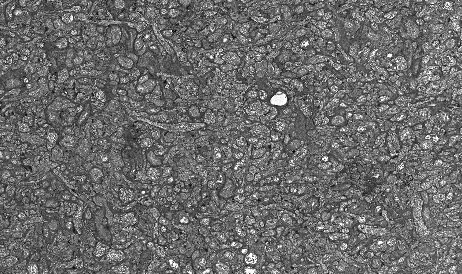
I am working on a paper that we are getting ready to submit with David Krizaj and his post-doc Peter Barabas when I got to looking at some of the retinal anatomy from this study. This dataset is part of a larger dataset that focuses on other areas of the retina, but this image composite really does exemplify how complex neural systems are and what we really take for granted when we see the world. This image (click on image or here to embiggen) shows the tangle of neuronal processes in the inner plexiform layer of retina that serves as the major retinal interconnection zone for many cell classes. There are hundreds, perhaps more, synapses in this particular image that serve as connection points between neurons, not to mention a large number of gap junctions that electrically couple subsets of neurons, all resulting in light detection and visual scene formation.
The analysis of this circuitry is one of the main missions of the Marc Lab for Connectomics whose datasets contain the most complete wiring diagrams of any complex neural system currently available. We look forward to continuing this work in ever bigger ways in the future.
This image is an transmission electron micrograph cropped down from a larger field and digitally mosaiced. This particular field has approximately 100 separate images out of a total of 700 images, captured at 5000x magnification.
