Dysmorphic Photoreceptors in a P23H Mutant Rhodopsin Model of Retinitis Pigmentosa Are Metabolically Active and Capable of Regenerating to Reverse Retinal Degeneration
Generation Of An Inbred Miniature Pig Model of Retinitis Pigmentosa
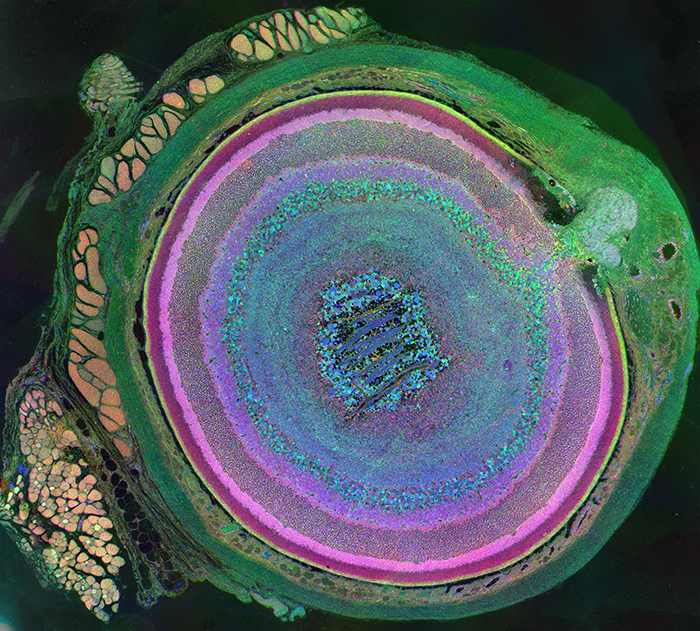
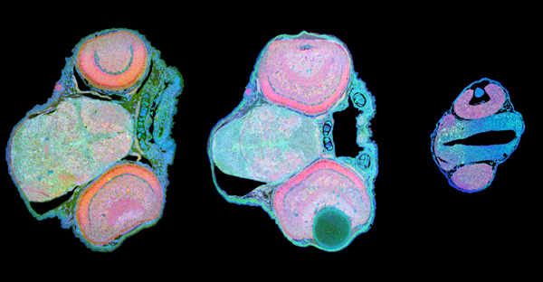
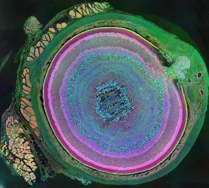
Metabolomic Eye Wins Science and Engineering Visualization Challenge
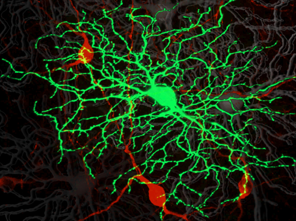
Developing Zebrafish Retina
Snowbird Neuroscience Symposium, 2011
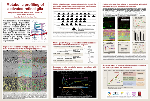
Metabolic Profiling of Activated Retinal Glia
Looking A Little Closer…
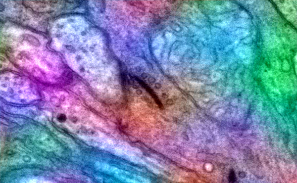
Ribbon Synapse In A Pathoconnectome
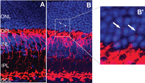
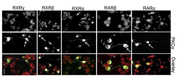
Retinoid Receptors Trigger Neuritogenesis in Retinal Degenerations
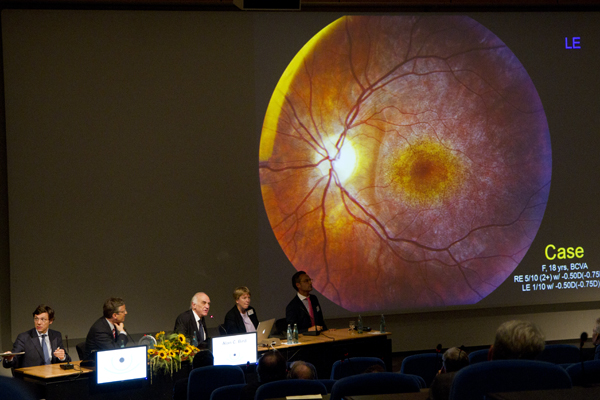
International Symposium on AMD 2011, Baden Baden
Metabolomic Eye
Retinal Remodeling in the Tg P347L Rabbit, a Large-Eye Model of Retinal Degeneration
Stargardt’s Disease angiogram
Venous phase fluorescein sodium angiogram of a dark choroid in Stargardt’s disease or fundus flavimaculatus. Stargart’s Disease is a progressive retinal degenerative disease caused by mutations in one of 3 genes, ABCA4, ELOVL4 or PROM1. The disease is a form of juvenile onset macular degeneration affecting the retinal pigmented epithelium (RPE). The disease onset and progression is …
Penetrating Corneal Injury Pigment
Photographer: Paula F. Morris, CRA, FOPS Moran Eye Center. FOPS diffuse slit lamp photograph of pigment growth following penetrating corneal laceration – 6 y/o patient. Image taken a using a Zeiss clinical slit-lamp biomicroscope with a Nikon D1S digital back at 16 mag. This image (#1 Alien) won First Place, Photo Slit Lamp Biomicrography Division, …