Science has been studying the retina for almost 130 years going back to the exquisite work of the great anatomist Santiago Ramón y Cajal in the late 1800’s. Building upon his work describing the structure of the neurons and glia, science has explored anatomy, physiology, genetics, biochemistry and psychophysics.
The more we study this delicate tissue, the more complexity we see. Our approaches for visualizing the metabolism of cells through computational molecular phenotyping (CMP), were developed to enable elucidating the structure and function of the retina and to reveal what goes wrong in blinding diseases of the retina like retinitis pigmentosa and age-related macular degeneration. To me, this work and the imagery that comes out of it is a constant reminder of why I work in retinal and visual sciences.
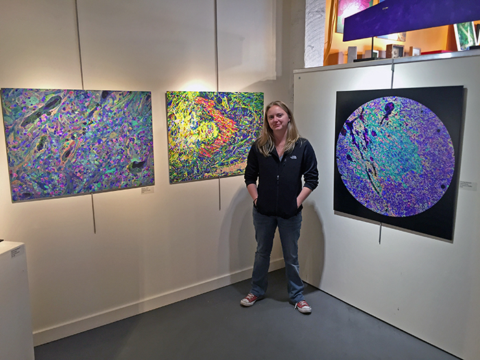
Getting the general public exposed to this work through compelling imagery communicates some of what we do in the lab to a wider audience and hopefully makes it a bit more accessible. So, when we recently had the opportunity to parter with the Moran Eye Center and Art Access to exhibit a scientific bio-image inspired gallery showing for the public, we enthusiastically started planning.
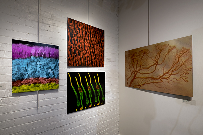
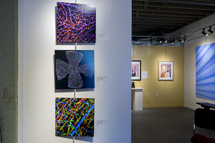
Art Access works with the disabled and disadvantaged to realize their artistic vision. The focus of this particular showing was on vision related art which played nicely with imagery derived from scientific analysis, but also art from the low vision or vision impaired community. The separate images shown above have been shown before and can be seen here (metabolomic retina), here (goldfish retina), here (classified retina), here (visual cortex neuron), and here (neuron pentaptych), but the exciting things about this exhibit was getting to see other scientists and grad students experience their first ever gallery showing as well as seeing people from outside science come to appreciate scientific visualization of something that many people hold near and dear, their vision. And it cannot be understated how powerful and empowering it can be for people to create art that represents their vision loss.
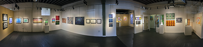
Participating artists in the main gallery with scientific images or scientifically inspired works included James Anderson, Michele Banks, Nico Cuenca, Jim Gillman, myself, Sarinda Jones, Helga Kolb, Gabe Luna, Paula Morris, Hope Morrison, Scott Peterson, Rebecca Pfeiffer, Stuart Stansbury and Peter Westenskow. Full resolution image of the above panorama is here (23MB).
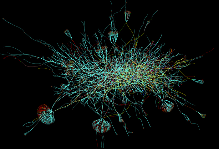
Image from James Anderson.
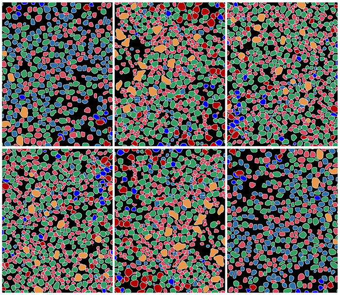
James Anderson contributed a stunning rendering of what we know about the connectivity of the retinal circuits. This is a mind blowing image that reflects a number of years of work and effort in elucidating how the retina is wired. Jamie’s artist statement on this work: “For five years the Marc lab has been collaboratively mapping the exact connections between neurons in the rabbit retina. This rendering of 5113 retinal neurons connected by 8631 synapses and gap junctions was manually entered by about a dozen expert annotators. While this representation of that effort as been stylized for appearances the same dataset has changed our understanding of how the retina processes information and how that processing may be altered by disease”.
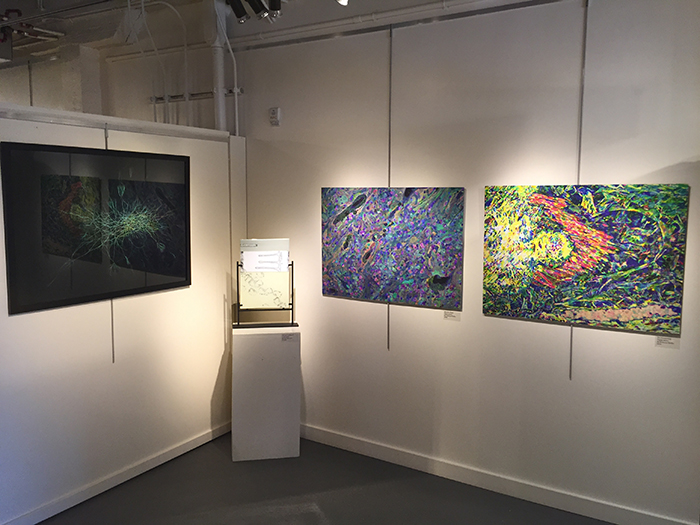
Rebecca Pfeiffer is a graduate student in the lab and contributed three works that derive from her exploration of Müller glia cells in the degenerate retina.
Glowing Astrocyte from Rebecca Pfeiffer
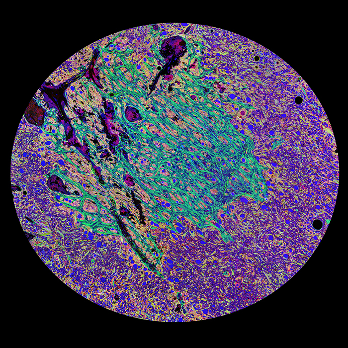
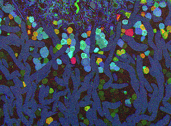
“This image is from the retina of a human who suffered from Retinitis Pigmentosa, a disease that destroys photoreceptors and eventually leads to blindness. The metabolic expression profile was obtained through a series of processing and staining steps, collectively termed Computational Metabolic Profiling (CMP). CMP is a powerful scientific technique allowing scientists to look at a wide range of molecules in cells or tissue from any organism on earth. The green structure is a specialized glia (neural support) cell called an astrocyte. These cells are crucial to the nervous system’s regulation of everything from metabolism to determining what to allow into the brain or retina from the blood. The impact of this cell(s) is made more pronounced by the elaboration of the arbors within a 5nm depth of tissue.”
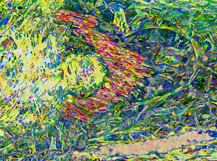
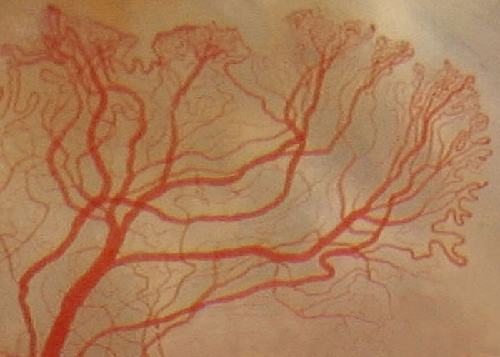
Red Boomerang from Rebecca Pfeiffer
This image is from the retina of a human who suffered from Retinitis Pigmentosa, a disease that destroys photoreceptors and eventually leads to blindness. The metabolic expression profile was obtained through a series of processing and staining steps, collectively termed Computational Metabolic Profiling (CMP). CMP is a powerful scientific technique allowing scientists to look at a wide range of molecules in cells or tissue from any organism on earth. The bright random background surrounding the focal red structure is the sclera of the eye. The sclera is the main supporting wall of the eye. The red structures are myelinated axons, which have traversed from the neurons of the back of the retina up into the sclera. Scientifically, this is incredibly interesting since this is never seen in the normal eye. This combination of the random components representing structure, and the defined nature of the axons demonstrating anomaly; is a cool dichotomy between science and visual impact.
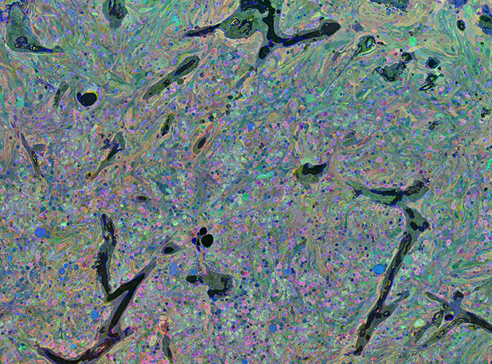
Metabolic Chaos from Rebecca Pfeiffer
“This image is from the retina of a human who suffered from Retinitis Pigmentosa, a disease that destroys photoreceptors and eventually leads to blindness. The metabolic expression profile was obtained through a series of processing and staining steps, collectively termed Computational Metabolic Profiling (CMP). CMP is a powerful scientific technique allowing scientists to look at a wide range of molecules in cells or tissue from any organism on earth. The swirling pattern that emerged in this image is allowing the visualization of levels of three molecules (taurine, glutamine, and glutamate) in neurons and glia of the eye. In essence, this image is science and crucial to the understanding of retinal disease, but out of the chaos of the disease comes beauty. Part of what made this image stand out in comparison to the hundreds of other metabolic profiles of diseased retina I’ve produced is the almost serenity that the image evokes, though in scientific terms the image is demonstration of quite the opposite.”
Blind from Hope Morrison
Hope’s statement on her amazing piece above: “Though art and science might be considered opposing interests, they are more related than mutually exclusive. Blind represents the marriage of these two pursuits. This image is retinal tissue labeled for multiple metabolites (taurine, glutamine and glutamate) using computational molecular phenotyping (CMP). CMP allows scientists to visualize the metabolic states of all cells within tissues simultaneously. This image is a particularly powerful example of the visual impact that scientific imaging can have. The beautiful color and clarity stands out in stark contrast to the reality that the tissue is diseased with Retinitis Pigmentosa, a degenerative eye condition that destroys photoreceptors and eventually leads to blindness”.
Artist Michele Banks (@artologica) Michele Banks is a painter and collage artist from Washington D.C. whose work is inspired by science. Her paintings have been exhibited at the AAAS and the NIH and featured on the covers of books and scientific journals. Her artwork has been featured by The Scientist, Scientific American, BoingBoing, and Wired, among others. She sells her work on Etsy, here and writes weekly about art and science at The Finch & Pea.
Michele’s artist’s statement is: “Over the past several years, I have created paintings exploring themes from science and medicine, including images such as viruses, bacteria, and dividing cells. Watercolor is an ideal medium for this type of work, because it can be transparent or translucent, allowing us to look at what is happening beneath the surface. It also naturally flows into fractal patterns such as those seen in the nervous and circulatory systems, as well as in tree branches and river systems – in fact, everywhere in nature. The structures of the eye, of course, follow these patterns also. And yet the eye – and its relationship with the brain – has an even deeper resonance for visual artists. Eyes and brains are our indispensable tools, and as such, an irresistible subject for artistic exploration.”
(I already bought the B&W ink retina image). Thank you Michele!
The above image shows art from Nico Cuenca on the left and Paula Morris on the right.
Paula’s artists statement was:
“Albert Einstein said, “Look deep into nature, and then you will understand everything better.”
As an ophthalmic imager, it has been my vocation to look deeply into nature; more specifically into the human body, to document patterns of health and of disease in the eye. I have been fascinated with the appearance of living tissue and often startled by the amazing colors and patterns I have seen through the lenses of the imaging instruments I use. Life seen at high magnification is extraordinary, and the in situ anterior segment images I have chosen to submit to Art Access I have re-identified as entities of another kind: beautiful, striking, and abstract.
Each of these images was made to document an ocular condition – in other words, pathology – an abnormal pattern. Each condition is remarkably different and yet each was located within the anterior segment of the eye, in structures that are within centimeters of each other.
An ophthalmologist once said of his practice, “That is what we do– recognize patterns. Normal patterns, and patterns of disease.” In these cases, I have interpreted the appearance of these abnormal patterns as images of other things in nature. In doing so, I believe I have found and emphasized the beauty in the image.”
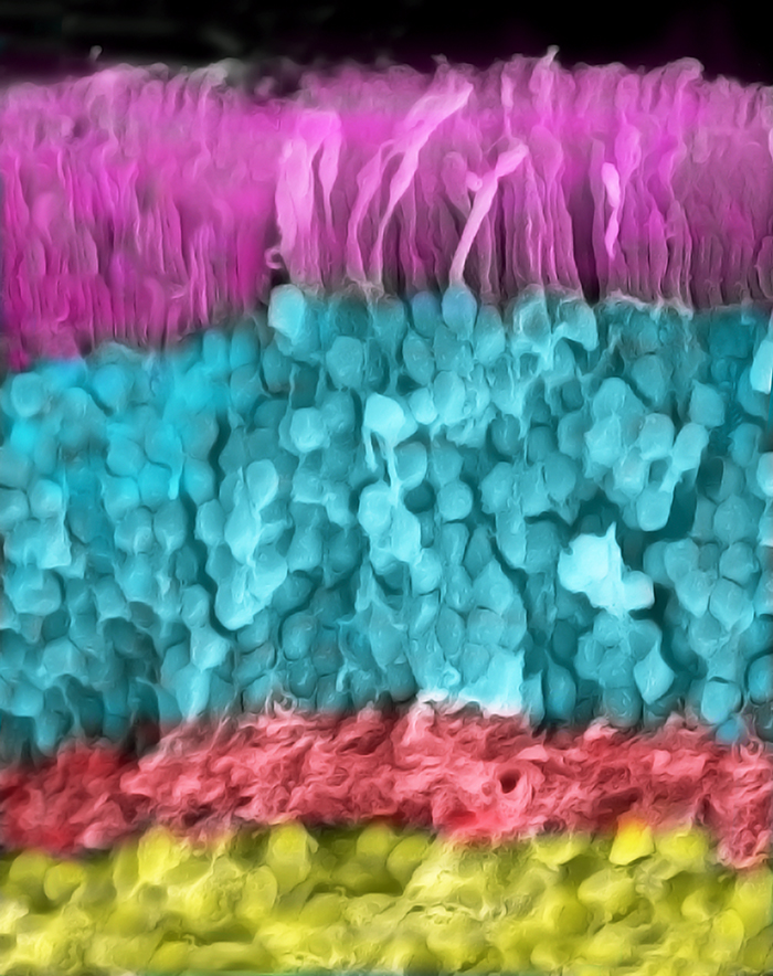
Image from Nico Cuenca.
Nicolás Cuenca maintains the excellent Retinal Microscopy website and has been a regular contributor of image and subject matter content to the Webvision site. His contributions to this show are remarkable and show everything from rat retina above to primate cones shown in the two images below.
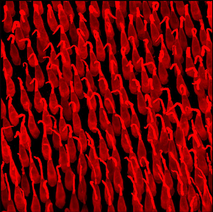
Image from Nico Cuenca
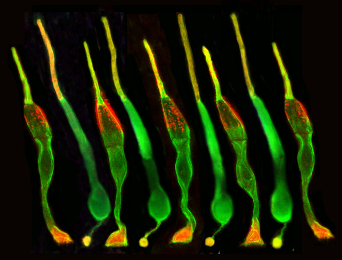
Image from Nico Cuenca
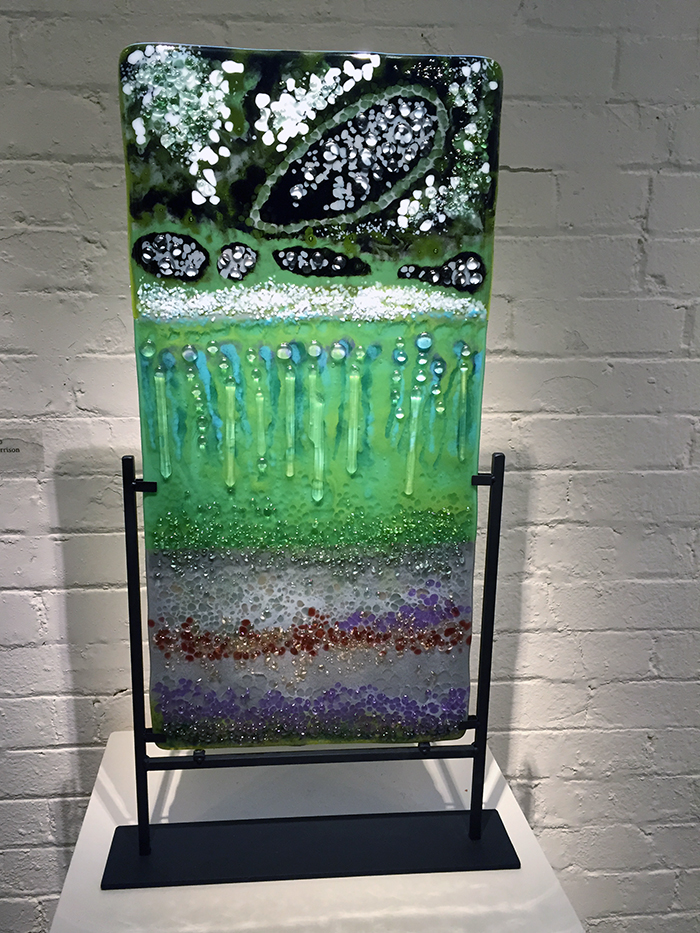
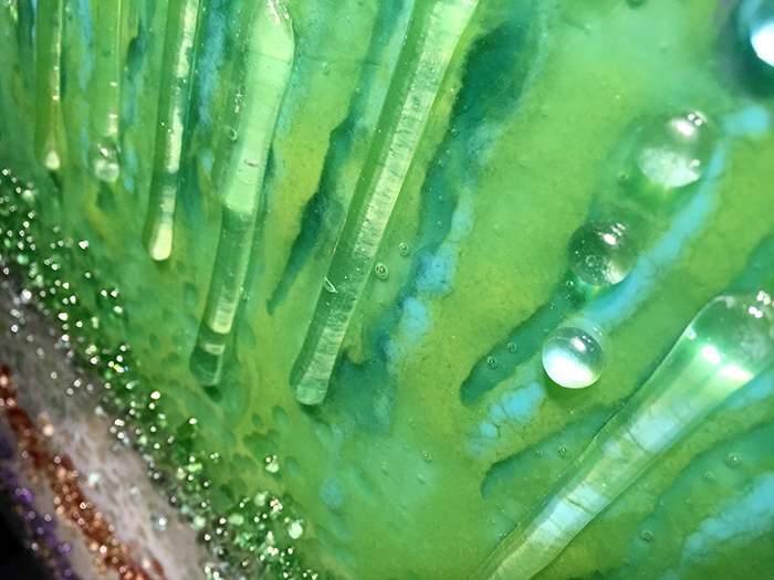
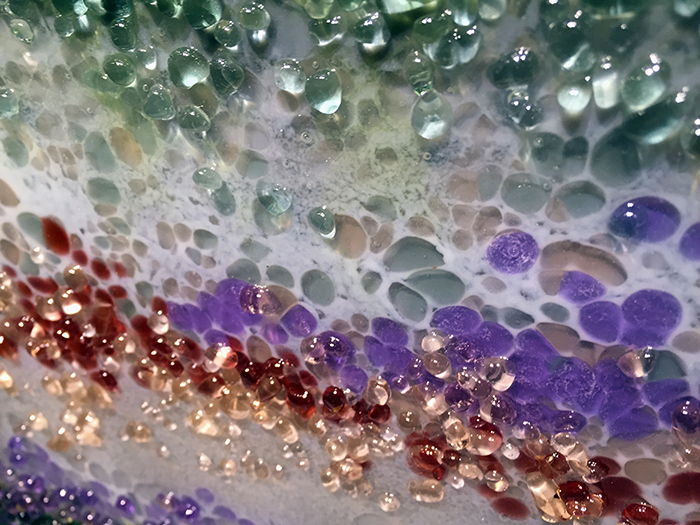
Glass artist Sarinda Jones made a series of amazing fused glass impressions of retina that really are stunning. She managed to capture the different layers of the retina with the ganglion cell layer, the amacrine and bipolar cell layer, the photoreceptors as well as the retinal pigment epithelium and choroid in this one piece. This piece was my favorite fused glass work of hers, though they were all spectacular representations. In others, she managed to stunningly capture some of the pathology seen in retina from patients with retinitis pigmentosa.
Gabe Luna is an award winning scientist from the lab of Steve Fisher and Geoff Lewis at UCSB sent a series of 5 amazing images he has created as part of his exploration of the retina.
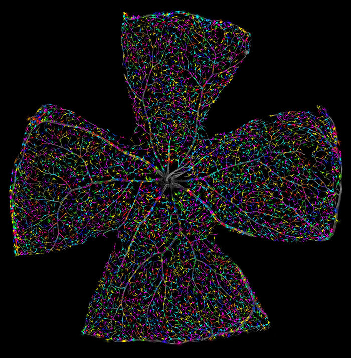
Image from Gabe Luna
Gabe writes: “The technicolor retina is an attempt to understand and characterize a largely enigmatic cell, the astrocyte. Using high-resolution light microscopy, genetic manipulation, and computational approaches to identify individual cells that provide new avenues for researchers to examine changes in the retina under conditions of development, disease, aging, or injury. Using these techniques scientists are able to extract morphometric data on a pan-retinal scale as well as maintaining the ability to interrogate structural changes at the single-cell level”.
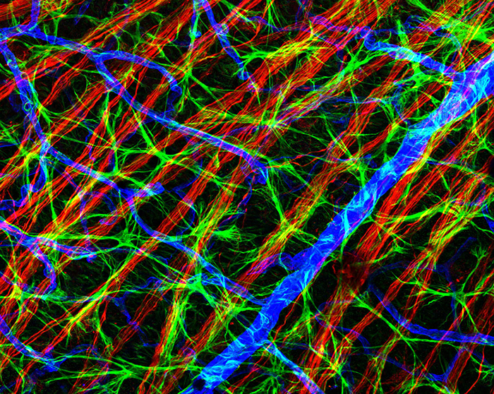
Image from Gabe Luna
Using an experimental model of retinal detachment to separate the underlying retinal-pigmented epithelium and the neural retina combined with immunocytochemistry and the use of confocal microscopy. Here researchers document the changes of astrocytic endfeet and their contacts with the vasculature and neurons. By capturing images at less than 1-micron intervals the resultant image is one that shows how the astrocytes begin to expand their coverage of the blood vessels in response to mechanical injury.
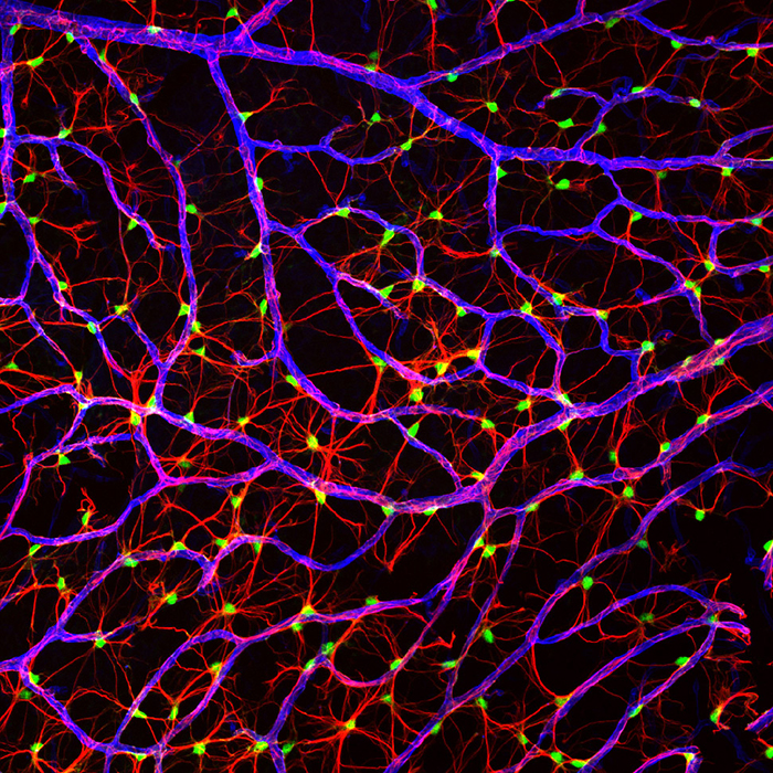
Image from Gabe Luna
Using genetic technology, specifically the green fluorescent protein individual astrocytes are easily identified, using this information, coordinates for individual astrocytes are noted and subsequently segmented by a random-walk computational algorithm. Using this methodology it spatial properties of retinal astrocytes are observed such as tiling or overlapping processes of neighboring cells, as well as amount of contact with blood vessels in close proximity.
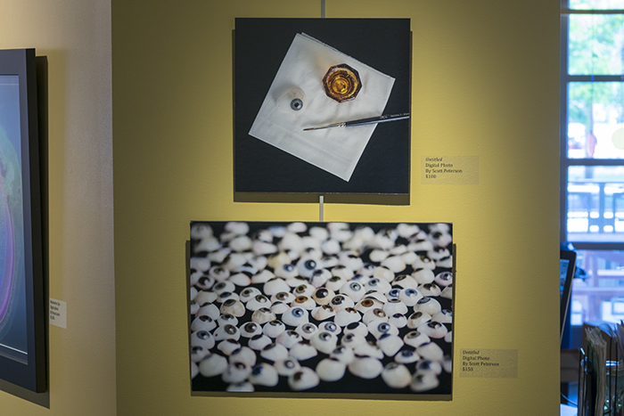
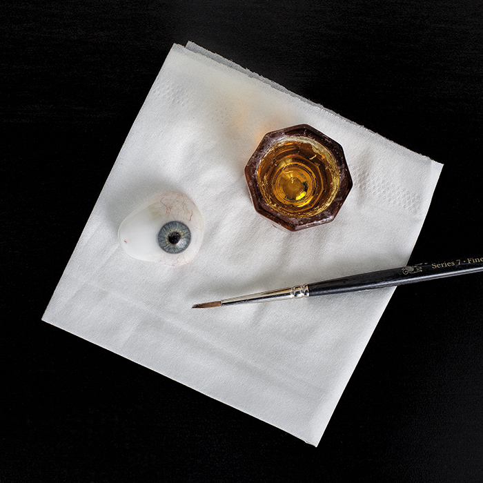
Image from Scott Peterson
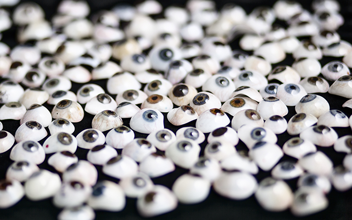
Image from Scott Peterson
Photographer Scott Peterson traveled out to visit an ocularist here in Salt Lake City to capture imagery of their work. Ocularists are specialists who create prosthetics for people who are missing eyes from trauma, disease or genetic conditions. Their work is amazing and Scott beautifully captured the sense of the work. His image with the paint brush is an amazing still life which has a very 16th century Dutch painting feel to me.
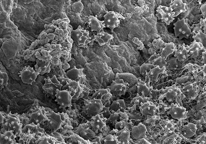
Image from Peter Westenskow
Peter Westenskow from the Scripps Institute in Marty Friedlander’s laboratory sent this scanning electron microscopy image of platelets from his work. It is always amazing to me how biology functions and something as insignificant as platelets are so critical to survival. You can clearly see the platelets and their activated state in this image which is a stunning representation.
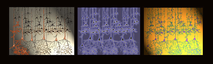
Image from Helga Kolb
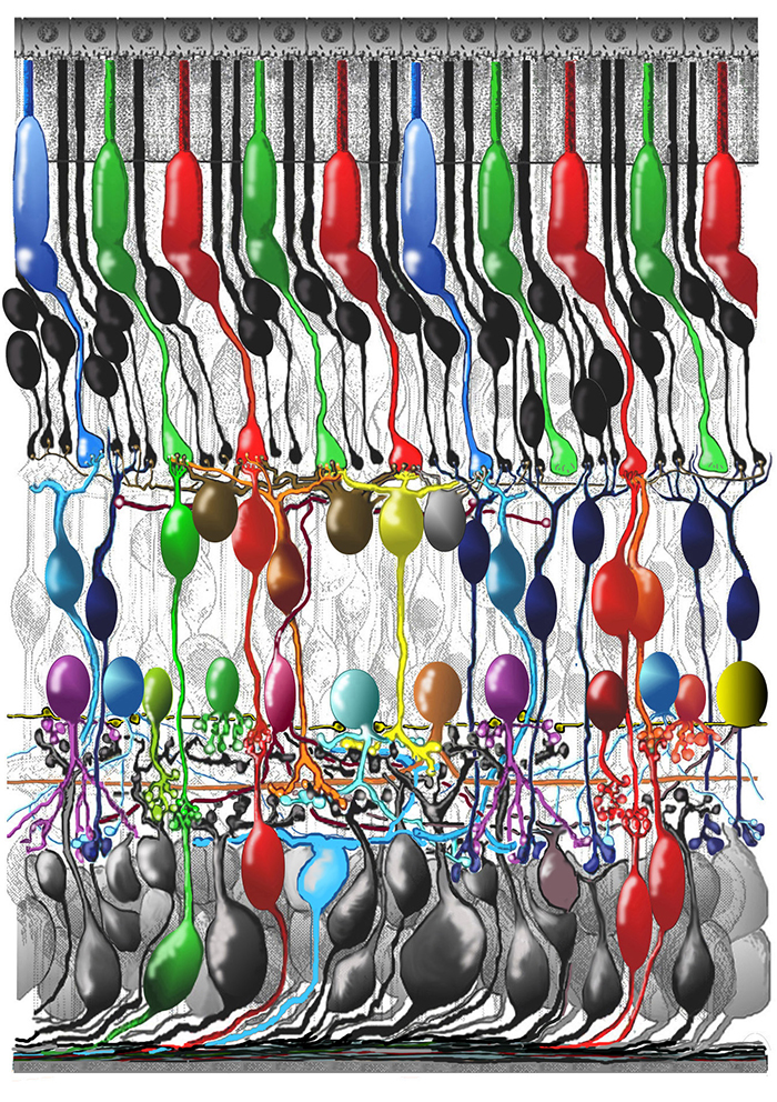
Image from Helga Kolb
Helga Kolb is an Emeritus Professor of Ophthalmology and Visual Sciences at the University of Utah where she worked for many years. Her work in visual sciences are landmarks in our understanding of retinal science and her contributions to the field cannot be underestimated. Additionally, Helga is the pioneer behind Webvision, perhaps the world’s first online textbook. Helga’s artists statement: “Helga Kolb is a self-taught painter who took up oil painting after retirement from a career in Science. She is presently an Emeritus professor of Ophthalmology at the University of Utah and decided upon retirement to follow the dream of her youth to be an artist. Helga’s professional work was in anatomical research on the retina and she found plenty of ways to express an artistic ability in that work. Light and electron microscopy are very visual research tools and the beautiful cells seen in the microscope need drawings and summary diagrams. In terms of oil paintings, Helga loves to paint the natural beauties of Southern Utah. She paints from photographs of rock formations in National parks, particularly Zion and Snow Canyons, Capitol Reef and Yellowstone. Paintings are large, mostly 24×30 and take off from the natural color of the rock formations into many hued bright colored fantasies. Helga has exhibited at the University of Utah, the Oasis Café gallery, Loge Gallery, the Jewish Community Center and in fund raising shows in Palm Springs, California. Her works are also in private hands, friends and family.
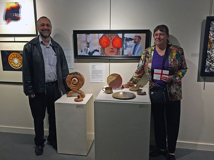
Moran Eye Center, ophthalmic imager James Gilman was also represented at the show with some stunning abstract art derived from his work in capturing images of the retina. You can see some of Jim’s ophthalmic imagery work here on Webvision. as well as many other low vision artists artists were represented at the show. Unfortunately, I don’t have permission to reproduce their images here, otherwise, I’d show you what a stunning assortment of images, mixed media painting, woodwork and photography they generated. One of the artists, Nancy Droubay is shown above on the right with Ophthalmologist Paul Bernstein from the Moran Eye Center just above.
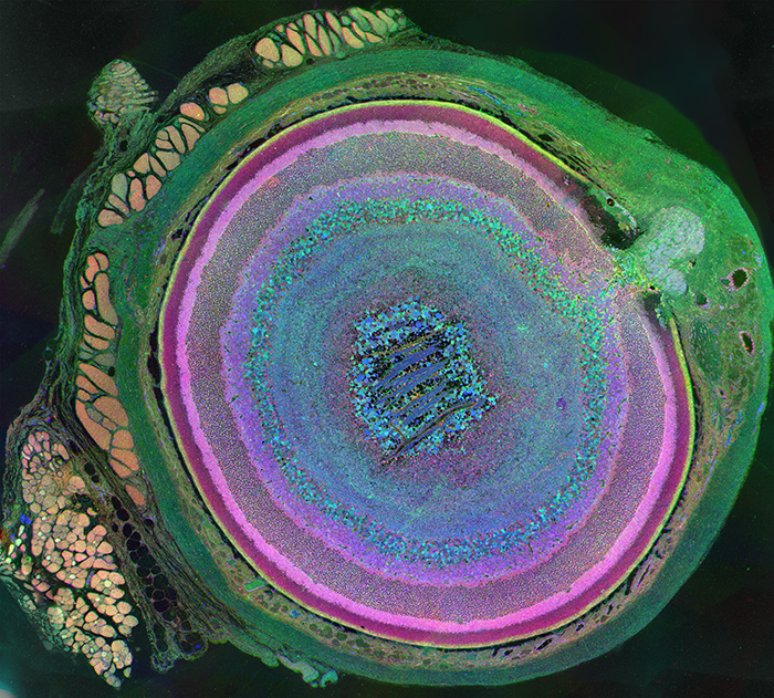
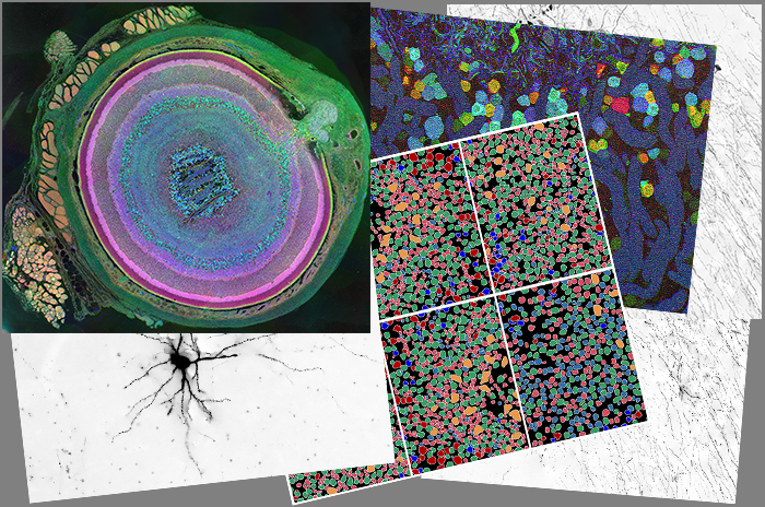
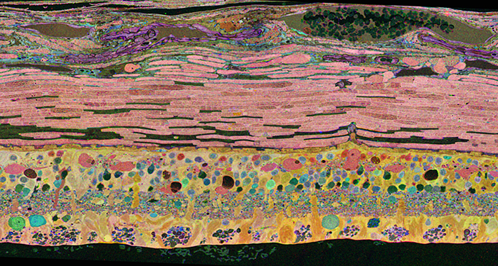
My famous Metabolomic Retina was on display. I have a writeup of it here.
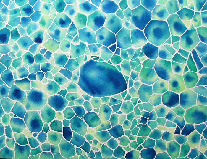
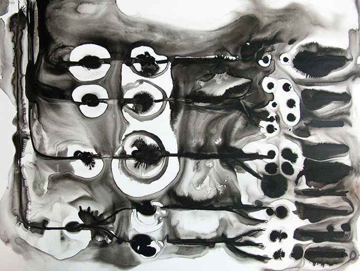
A former Christmas card that we’ve turned into a full wall panel at a couple of installations was also on display.
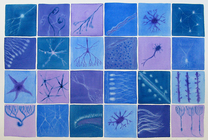
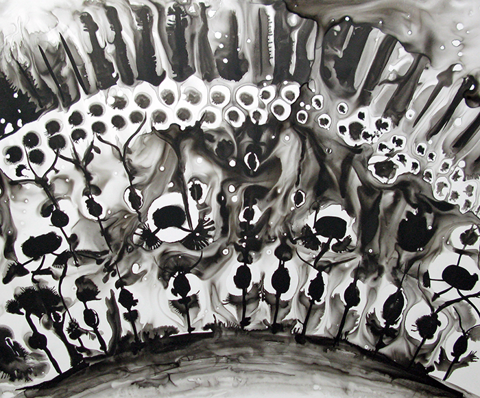
A triptych made up of some of these images was also on display. It originally appeared here.

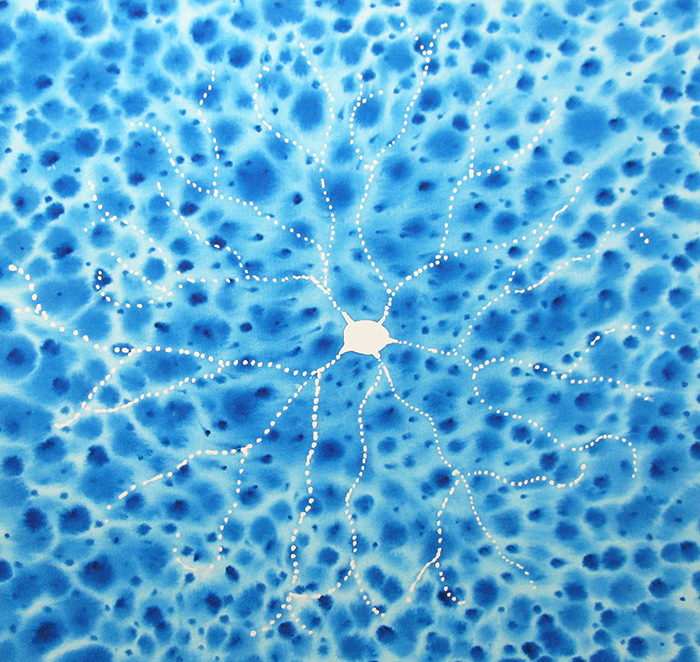
And I had an image of a cortical neuron on display that always reminded me of a pencil or charcoal drawing on paper. It originally appeared here.
And finally we had a FASEB Image Award winner on display with an explainer on the image that appeared here.
Many thanks to City Weekly for the shoutout on the show and to The Finch&Pea.
Beautiful, beautiful blog!
:-) Thank you. All the images from the artists make it easy on this post.
I’m glad I don’t have to be a judge or pick a favorite. Great collection of art. Thanks for posting it.
Thank you Bill. It was quite a bit of fun working with everyone on this.