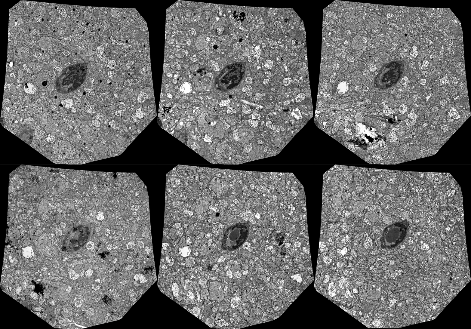
I was going through the archives of really old image data after a conversation with Liz Jurrus about some new work and came across this dataset. It looks rough with a little dirt on it in places, but this was one of the *very first* datasets a number of years ago where we took a group of transmission electron micrographs, mosaicked them together and then registered each slice to one another to allow us to move through the volume. What you are seeing are 20 registered frames through a dataset from mouse retina following a blood vessel. This is old technology right now, and there are a number of groups around the world that are doing this, some with tools we helped build with SCI, but this dataset was the very first proof of principle that these technologies would work in our hands, allowing us to create frameworks to understand neural circuitry, then go on to reconstruct entire neural circuits.

Thanks for the interesting image Bryan.
I thought you might like to see and article about cockroach night vision on the imaging resource web site. Since you are a photographer you may have already seen it.
http://www.imaging-resource.com/news/2015/03/16/scientists-discover-cockroaches-use-a-long-exposure-photography-technique-t
Yeah, I’ve seen that in a number of places now. Very cool and an indicator of systems that integrate overall luminance. There are systems in the mammalian retina that do the same, reporting to the supra chiasmatic nucleus (SCN) in the brain to set the circadian rhythm.
That sounds absolutely amazing. Can you explain how that happens in a little more detail. Is there a known mechanism to account for how those systems record light over a period of time?
Yeah, you can check out a chapter on melanopsin ganglion cells over on Webvision: http://webvision.med.utah.edu/book/part-ii-anatomy-and-physiology-of-the-retina/elanopsin-ganglion-cells-a-bit-of-fly-in-the-mammalian-eye/
Thanks for the link Bryan. I was not aware that ganglion cells could also be photoreceptive. The various interactions and feedback mechanisms within the retina and brain are truly mind boggling.