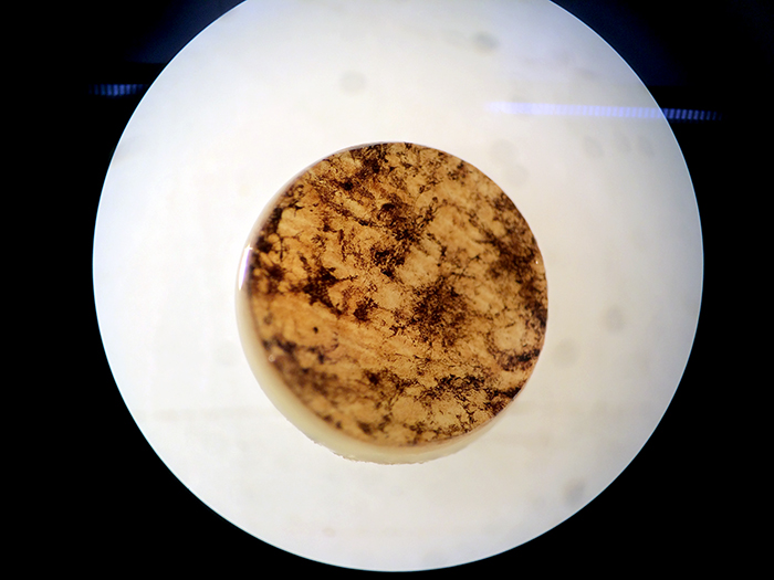
We are working in the lab today with retinas from patients who have donated their eyes to science after they have died. This portion of retina is a 4mm punch from the eye of a patient with retinitis pigmentosa, a disease we study that alters the topology and circuitry of the retina during and after photoreceptors degenerate.
I’ve talked about this a little bit before, but the clumps of dark pigment in this image are abnormal and are clinically called pigmented bone spicules which are formed from a single layer of cells called the retinal pigment epithelium that is getting pulled down into the neural retina, forming clumps of pigment. What you can’t see from this image is all of the histological changes in the tissues and the neural circuitry changes that forms the basis for our work. More to come…
We are grateful for those who donate tissues and organs to research and endeavor to learn everything we can from these donations. We could not do what we do without the generosity of those who donate their bodies to science after they have passed.
Wow, thanks for all your hard work Bryan.
It is what we do Laura. Just happy that we can continue doing the work.