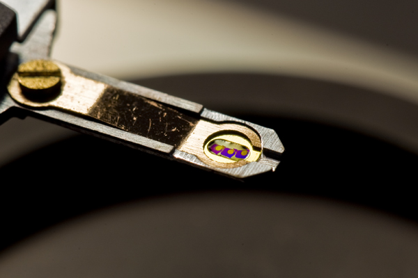
Small is relative… I put this image in the small life category because this is a very small thing you are looking at and it was taken with a macro lens, but there are things much smaller still.
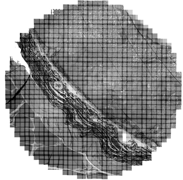
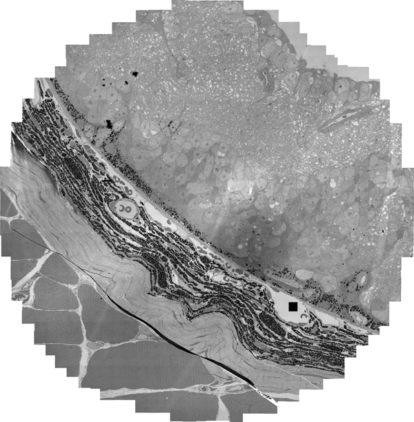
To give a sense of scale on the retinal reconstruction project we are working on, I am showing you a holder for EM grids. This device is inserted into an electron microscope and the sections of tissue you see in this image (the three little purple squares) are then imaged. Each one of the little circles in the purple tissue section isthe area that is actually imaged by our JEOL JEM-1400 electron microscope with our custom software and hardware assembly to create images that you see below. The images below are examples of dramatically subsampled image from an original image size of 30GB. The first image shows the mosaic of images that comprise the overall image and the second image has the mosaic lines removed.