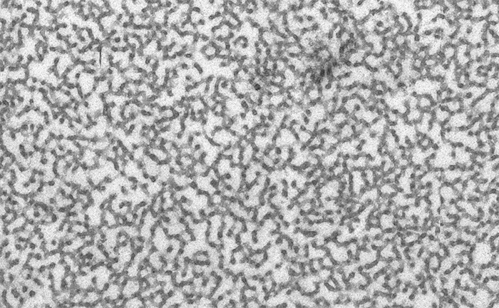
I’m busy preparing a manuscript that describes the changes in the retina of patients with a degenerative disease called retinitis pigmentosa that leads to massive alterations in structure and circuitry. As part of that, I’ve been spending quite a bit of time with ultrastructural data and came across the following field with regularly repeating texture. I’m not actually sure what this is to be honest, but it came from within a vascular component from the retina of a person with retinitis pigmentosa.
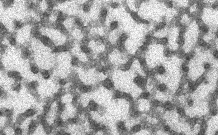
Each one of these tubular like structures seen in the zoomed in image above has an incredibly regular 34nm diameter. Is it a lysed cell? Cellular components that have polymerized? I don’t rightly know…
WHERE in the retina (wrt to fovea and also what layer?)
Aha! Mid periphery, temporal retina. As to what layer? Good question given that the topology is so far gone…