
Progression of Geographic Atrophy
These retinal images are from an 84 year old white male who presented to the Moran Eye Center in 2008. He was diagnosed and followed for dry age-related macular degeneration (AMD) with serial autofluorescent photographs showing progression of geographic atrophy of the RPE from 2008 to 2014. These images were prepared by James Gilman of the Moran Eye Center.
Flying from Charles de Gaulle To Chicago, A Time-Lapse

Retinal Astrocytic Hamartoma
This is a 58 year old white female with a retinal astrocytic hamartomaon her right optic nerve. Retinal astrocytic hamartomas are glial tumors of the retinal nerve fiber layer arising from retinal astrocytes. This animated GIF file illustrates the height of the hamartoma and is another example of where animated gifs can be a fantastic teaching …
Failed Experiment
Museums Of Paris

Paris In The Rain

Portrait: Sawyer Pangborn (In My Office)

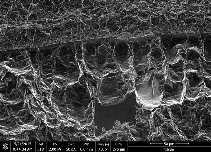
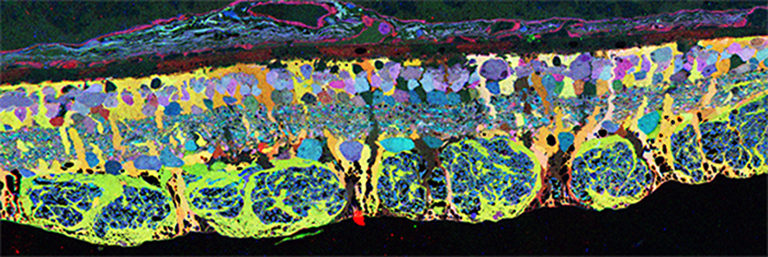
Müller Cell Metabolic Chaos During Degeneration
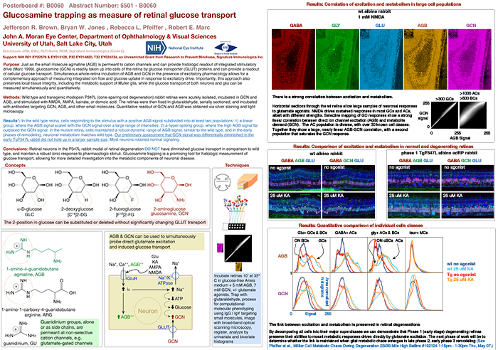
Glucosamine Trapping As A Measure Of Retinal Glucose Transport
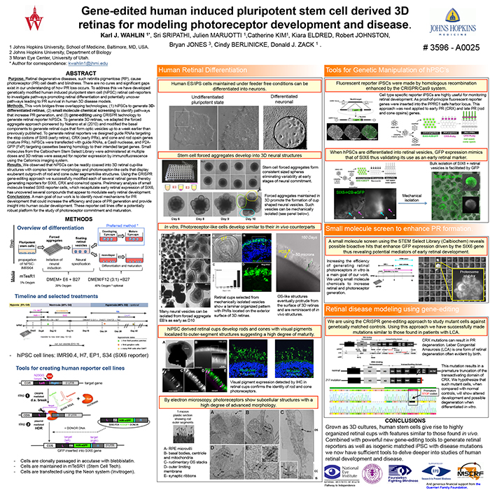
Gene-Edited Human Induced Pluripotent Stem Cell Derived 3D Retinas For Modeling Photoreceptor Development And Disease
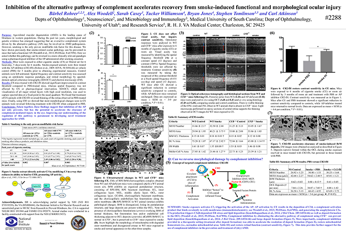
Inhibition Of The Alternative pathway Of Complement Accelerates Recovery From Smoke-Induced Functional And Morphological Ocular Injury
Portrait: Poppy The Cat