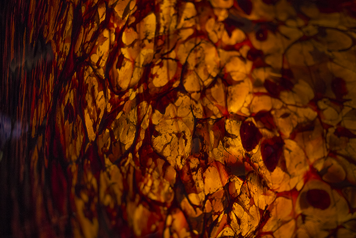
In yet another example of art imitating life, I was going through some imagery from earlier in the year and came across this image of blown glass up at the Chihuly garden up in Seattle which stunningly, reminded me of what the human retina looks like in a patient suffering from the blinding disease, retinitis pigmentosa (below).
I would be very curious to know just how this glass was formed…
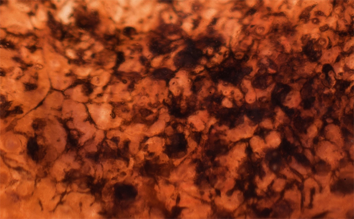
This is an extreme closeup image that is backlit and magnified of the inside of a human eye showing the retina from a patient suffering from retinitis pigmentosa. This image is focus stacked to give an enhanced depth of focus to illustrate the structure of those dark structures called “pigmented bone spicules” in an intact globe. The pigmented bone spicules are remnants of the retinal pigment epithelium that have been dragged down into the retina as the retina has undergone retinal remodeling, a process that happens when the photoreceptors degenerate in blinding diseases. A version of this image appeared in our last review of retinal remodeling and is precisely what I thought of when looking at the piece of Chihuly glass.
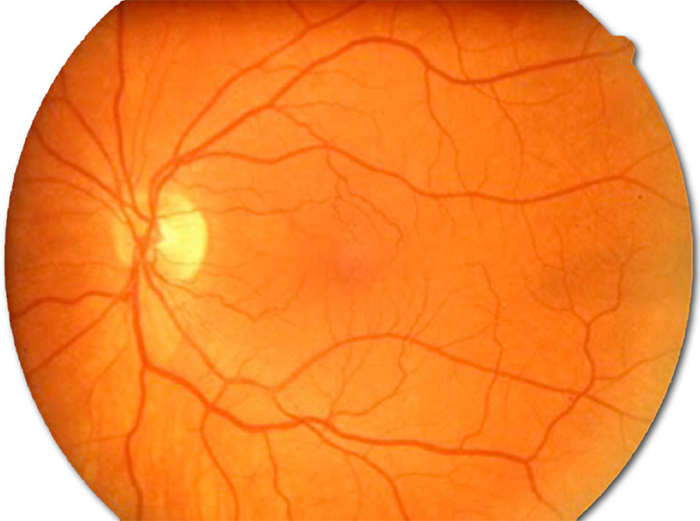
For some context, this is a wider view of a normal human retina as imaged with a fundoscope. Normally, the retinas appearance should be more smooth with uniform color and blood vessels are smooth and not tortuous. The retina is an out pouching of the brain that has all of the cells and circuitry in it to capture photons and start to structure them in a manner that allows us to visually perceive the world around us.
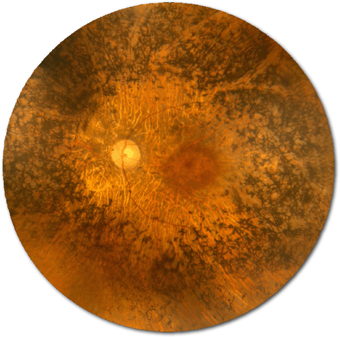
This is also a human retina imaged with a fundoscope. You can see that things look very different late in the process of retinitis pigmentosa after retinal remodeling is advanced. The pigment clumps up into those pigmented bone spicules, the blood vessels are narrower and more tortuous and the smooth yellow/orange/red color of the retina is much more mottled.
I’d love to see some glass that specifically represents neural anatomy and the retina. We have a huge space in the Moran Eye Center lobby that could be an amazing installation site for large glass art dedicated to the eye or retina. Imagine what a “glass” retina would look like in a 60 foot tall space…
That’s quite the difference in the two eye pictures. It looks like it’s worth the further you are from the center. Does that mean the vision is worse away from the center as well. What would it be like? Blurred or does one actually see darker there? What causes this?
In retinitis pigmentosa, people go blind from the periphery in towards the center of their vision. It is the opposite of what happens in age related macular degeneration where people go blind from the center, out. As far as causes, it turns out there are many hundreds of reasons why this happens. From gene defects in protein processing and trafficking to defects in rhodopsin packaging and processing as well as many others. In short, anything that stresses the photoreceptors to the point where they die results in this process.
it’s amazing how much they look alike. very cool.
Seriously… I was kind of blown away when I saw that glass work.
The same person could have retinitis pigmentosa, color blindness, and cystoid macular edema. Is a fundoscope capable of showing details to the level that researchers could recognize that more than retinitis pigmentosa is involved? What is the best available gene therapy today?
This is true. All could co-exist in the same eye. The fundoscope would not be able to determine color blindness.
Concerning gene therapy, there have been over 1800 gene therapy trials of all kinds (cardiovascular, cancer, cyctic fibrosis, etc…) world wide and a few in vision science including some in choroidermia, Stargardt’s disease, autosomal recessive retinitis pigmentosa, Usher’s syndrome and wet AMD. These are all in early stages and the focus has been on safety, though some folks are reporting improvements in vision. That said, those reports are not well defined yet and days are very early. Much basic science needs to be performed still as much of the gene therapy trials (if not all) are being done without a knowledge of the tissue and circuit level impact in my opinion.
We have had a couple of trials here in animal models and are currently chasing some of the more precise biology behind them, but are not doing any clinical trials in gene therapy ourselves.
That said, there is good science out there waiting to be done, but science cutbacks and the government shutdown have slowed or halted quite a bit of vision science research. What can you do about this? Contact your senators and representatives and express your feelings on the issue. People are going blind. Scientists can and want to do the work. They need funding to be able to continue chasing the goals of curing blinding diseases.