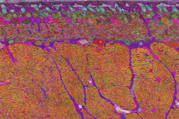
For Halloween, we have a scary, vertical section of retinal Computational Molecular Phenotyping (CMP) data in seasonal colors of a model of degenerative retinal disease near the central part of the retina. The thickest portion of the image at the bottom are the axons from ganglion cells as they stream out of retina into the optic nerve. The purple streaks in-between them are the Müller cells one of the glial cells of the retina. Above the fiber cell layer is the ganglion cell layer with the output cells of the retina. The inner plexiform layer or principle connectivity layer of the retina is the next layer, though we are finding that there is extensive connectivity throughout the retinal layers including the fiber cell layer. Above that is the amacrine cell layer, bipolar cell layer and the remnant photoreceptors in cyan just below the retinal pigment epithelium at the very top.
